
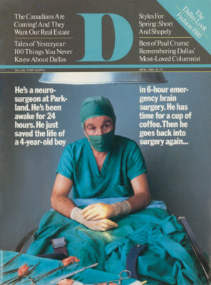
Saturday night. Hunt Batjer is doing what he always does on Saturday nights: waiting for someone to get hurt. Sooner or later the call will come: a .32 slug through the temple, an ice pick through the spinal cord, a forehead through the windshield. It never fails. Some accident out there looking for a place to happen will find it. Some poor slob minding his own business will wind up in the wrong place at the wrong
time. On Saturday night, especially on Saturday night, none of the rules apply. You can get killed just for being alive.
Hunt used to hate the Saturday nights. The endless stream of opened heads and cracked skulls, broken backs and seizures and strokes; the whole crazy scene in the Pit angered him. He didn’t like the idea that he was at the mercy of some drunk on a highway or some pulsating aneurism that just decided to blow. But three years of neuro-surgical residency – 154 Saturday nights, to be exact – have beaten the rebellious streak out of him. Saturday nights are just part of the program. So you want to be a neurosurgeon? Okay: We’ll teach you all about meningiomas and metastases and subdural clots; we’ll teach you how to suture a laceration, how to gut a tumor, how to beat an aneurism to the punch; we’ll teach you how to tell a mother her son will never read or an athlete why he won’t walk again. But mainly we’ll teach you how to get through the Saturday nights.
That is the point: Getting through. That is the bottom line of residency, the final measure of whether you have what it takes. Half a dozen gunshot wounds, a stroke or two, a battered spinal cord – you get through it. You learn a lot on emergency surgery, down in the Pit: how to work four, five, ten hours on a patient, knowing he’s going to die anyway; how to stay alert even though you’ve been up for 24 hours. The Pit teaches you patience.
While waiting for the emergency beep, Hunt hustles about the wards at Children’s Medical Center, tending to chores that cropped up during the afternoon. There is a shot to be administered here, a discharge paper to be filled out there, a watered brain to be tapped, a nurse to be reminded, a mother to be reassured. More stuff to get through.
The call comes early, about 7 p.m. A nasty wreck on a country road outside of town. The victims: the male driver, unscathed except for bruises; a young female passenger, who has suffered massive and multiple internal injuries, as has the eight-month-old fetus she carries in her belly; a two-year-old boy who hurtled headfirst into the dashboard on impact. Hunt drops his paperwork and races down the bright corridors of Children’s into the dank, shadowy halls of Parkland, past the accounting department, the ob-gyn clinic, the all-night pharmacy, until he reaches the familiar sign, “Surgery.” The Pit is a scene from Fellini: The broad aisles are choked with stretchers bearing the bawling young, the comatose elderly, all the shapes, sizes, and colors of people who have wound up on the wrong end of an accident. Scattered among them, seated in hard plastic chairs, are the “lucky” ones: the broken arms and battered heads, the toothaches and Saturday night nosebleeds. Residents, interns, and nurses, in surgical green and blue and white, ask questions and bark orders, give shots and take blood pressures, suture wounds and check dilated pupils, all in a spastic ballet.
Hunt checks in with the “pit boss,” who briefs him on the case. General surgery in Emergency will handle the woman, who is fading fast from pulmonary trauma. As the resident in pediatric neurosurgery, Hunt will take charge of the two-year-old. The boy is in Trauma Room 1, which is never good news. The Pit has six main trauma rooms; the current load of patients is assigned and shuttled according to severity of injury. Anybody in Trauma Room 1 is seriously hurt.
Hunt’s fears are confirmed: The infant was thrown into the dashboard so violently that his forehead shattered; it just isn’t there anymore. The underlying tissue, including the dura – the taut, leathery “glove” that covers the cerebral cortex – is badly shredded. In the rear of the boy’s skull, a massive laceration stretches from ear to ear. It’s gushing blood. Already his blood pressure is nearing SO, indicating he is going into shock. It’s a neurosurgeon’s nightmare: brain oozing out of one end of the head like toothpaste, blood pouring out of the other end like water from a broken main. It looks like an occasion for one of the Pit’s blackest jokes: “He’s going to die. Let’s just take a look here and see what’s going to kill him.”
Hunt orders that the laceration be pressured and cleaned, to thwart immediate threats of hemorrhage and infection. He asks for transfusions and placement of an inter-tracheal tube, by which the child can be hyperventilated to reduce swelling of the exposed cortex. He checks the infant’s sag-gital sinus to be certain it isn’t violated; a split there, and the child will bleed to death in a matter of minutes. Meanwhile, general surgery completes its evaluation of the rest of the body. With the exception of his shattered skull, the boy’s bones are intact; his kidney and liver functions are good; his lungs are solid; there is no apparent internal bleeding. It’s a neurosurgical problem, pure and simple.
As the boy is “toned up,” Hunt runs a cursory check of his neurological vital signs. Surprisingly, the child is at least semi-purposeful, with reflexive movement in his arms and legs and the all-important “doll’s eye” reaction when his head is twisted to and fro. He is actually somewhat lucky: The damage is confined to his temporal lobes. The boy has effectively been given a loboto-my by the dashboard, and aspects of his personality will be altered as a result, but the more crucial areas – sight, hearing, speech, and involuntary muscular movements, such as breathing and heartbeat – are apparently untouched.
Hunt has a decision to make. He can keep the child alive for the time being, but surgical strategy is not at all clear. He has two options. He can quickly cauterize and suture the laceration and whisk the boy up for a CT scan, a set of serial X-rays. That might tell him whether the victim has serious hemorrhaging in some part of his brain not immediately visible. A severe trauma in one region can set off a chain reaction of traumas in other areas; often the surgeon can never really be sure when they’ll erupt without a CT scan.
But a scan will eat up precious time. Since a large portion of the boy’s cortex is exposed, a special plaster cast will have to be sized and cut to protect his frontal lobes from radiation. That might take an hour. The scan will take another. Two hours, with the boy descending into shock. The boy might arrest while the film is being developed. Hunt intuitively knows the best way to get through it: He’ll put the boy under the knife first, scan him later.
Hunt calls his boss, the University of Texas Health Science Center’s chief of pedi-atric neurosurgery, Fred Sklar, for a second opinion. You always get a second opinion. There is no room for heroics, not when a two-year-old child lies there spilling his brains and his blood. Sklar listens carefully to the breakdown of the case and quickly agrees that Hunt’s approach is correct.
Hunt sets about the task of stopping the bleeding from the back of the boy’s skull. It is a slow and painstaking process, one that medical science still has no short cut for. You just have to stay with it, repeating the same treatments over and over. Hunt uses the typical combination of hemostatic treatments: A bipolar coagulator, a delicate set of pincers that emit a mild electric shock which immediately cauterizes the flesh; tiny strips of cottonoid and gauze; a small “sucker” to eliminate any stray blood the other treatments don’t catch. Satisfied that the internal bleeding is stopped, he quickly sutures the epidermis from ear to ear, his large, athletic hands moving with the speed and precision of a seamstress.
He rolls the boy over and goes to work on the messy anterior wound. After clipping away splintered bone, Hunt stops superficial bleeding, then trims away damaged portions of brain. He works efficiently and routinely, cutting out pieces of the boy’s brain. It’s simple: Pulped, lacerated, torn brain is dead; it isn’t working and it never will again. You get rid of it.
Hunt checks his watch – 2 a.m. – and moves to the most difficult task of all. Things are cleaned up, but there is sure to be extensive bleeding in interior portions of the brain. With a head shot like this there could be small hemorrhages, mounting clots, edemas almost anywhere. He has to go in.
He checks for signs of errant blood and makes the appropriate incisions, then opens them gently with a brain retractor. There is bleeding, all right. Hunt goes to work with the bipolars and the sucker, eliminating any trace of leaking on the inside. Satisfied, he irrigates the wounds and begins the process of closing up. That is no “easy ride home.” The fact is that the boy has no dura. Without it, cerebrospinal fluid could swell and crush the brain. The only solution is to patch in a makeshift dura. He slices a sheath of periosteum, the sinewy tissue that covers bone, from another portion of the child’s skull, sizes and trims it like a tailor, and begins stitching it to the remaining dura.
He works mechanically, each stitch matching the others precisely. It follows a concept basic to the surgeon’s craft: You do the same things every time. By 7 a.m. Sunday he is finished. Both ends of the boy’s head are closed. He is intact. His vitals are fair, his neurologic responses still encouraging. He is alive. He will undoubtedly have an altered personality, maybe some learning deficiencies. But what started as a pulpy, bloody mess is a sutured, breathing body. Hunt doesn’t feel proud or happy, just tired and a bit relieved. He’s gotten through, too.
The infant is “wired” in the intensive care unit at Parkland, and Hunt is finally free to go home. For a shower, at least. He gets in his green Datsun, drives down Harry Hines, up Wycliff, down Lemmon to Central, and on home to the Village. He drops his keys on the bar, shucks his clothes, and climbs into the shower. He stands a long time under the scalding spray, letting his thoughts wander, thinking about anything but brain surgery.
Hunt Batjer is tall, muscular, handsome. His thick black hair is stylishly overgrown and he sports a drooping mustache. He looks like a professional athlete, which is what he wanted to be before he got hooked on axons and the somatic sensory cortex.
As far as he is concerned, it amounts to much the same thing. Major league pitching or major league brain surgery. Two outs, the bases loaded, the three-two pitch; the eleventh hour of the operation, the patient’s blood pressure unstable, a big, angry aneurism ready to blow up in your face. Either way, you have to act. If you have good stuff, you pull it off. You win.
Hunt loves to win. Competition is in his blood: His mother is a noted musician, his sister a rising one, his father a successful insurance company executive in San Angelo. He was raised to be an over-achiever. He decided to become a brain surgeon because, next to baseball, it was the most competitive thing he could find. With abandon he plunged into the study-eat-sleep-study grind of pre-med, ignoring his peers in the Baby Boom Generation, who took cinch electives and pursued what he considered vague degrees in sociology and English. Hunt spent long nights in the library and the lab. To him, college was no joke, no four-year junket to “get your head together.” Getting through became a daily thing.
In med school, he disciplined himself to become a recluse; it was the only way to beat the system. Southwest Medical School in Dallas was ferociously competitive, one of the top five medical schools in the country. Several of his acquaintances decided early on that they would just survive the four-year ordeal, return to their home towns, and hang a general practice shingle. For them, med school was probably fun. They took it relatively easy, went on dates, drank beer in the afternoon, took weekend trips home. That was fine if you wanted to be a doctor. Hunt wanted to be a brain surgeon.
When the four years at Southwest Medical were through, he and 700 other aspiring surgeons applied for residencies at Parkland. Twenty-seven were selected for all specialties. In neurosurgery, a small unit with a growing national reputation, there was one slot. Hunt filled it. He loved the competitive simplicity of it: There was no room for hedging or rationalizing. Either you were it or you weren’t.
Being it was vital to him. A residency in thoracic or orthopedic or cardio-vascular wouldn’t have been enough. Hunt had no interest in fusing shattered bone or clearing out cancer-ravaged intestines or even making a clogged heart work again. The brain was it, the last mystery organ where a surgeon could never feel safe going in. It was beautiful, physically beautiful: glistening, clean and white, laced with an intricate network of blue and red strands; so simple, so complex. When a brain surgeon went in, he wasn’t looking at the body; he was looking at the person. When the patient came out of anesthesia, it was something religious: I just saw where your words and thoughts and feelings come from.
Monday morning. It is 6 a.m. and Hunt Batjer is just starting his morning rounds at Parkland and Children’s. He makes them at his usual breakneck clip, long legs striding over the green linoleum floors. He stops to check on a young girl who was hit by a car a few days before. The blow fractured her left parietal bone, the side of the skull. Though still semiconscious, she sustained no worrisome internal injuries. Still, she bears careful watching: There is nerve damage on the left side of her face and she isn’t yet speaking. She moves her limbs, but barely responds when asked to make a fist with her right hand. A sharp blow to the head essentially causes a short circuit in the brain’s intricate system of electrical impulses: Power remains, but its normal routes of conduction are scrambled. With no enduring bleeding or other problems, the patient can usually “gut” his way out of it – just heal himself. But you can never tell.
Hunt checks the girl’s neurological vitals by pressing and tapping at the major reflex centers: elbow, knee, ankle. As sophisticated as neurosurgery has become, the basic tests still tell you most of what you want to know. This morning, the 10-year-old is showing marked improvement. She is still “out,” but she has regained tone in her reflexes and her garbled moaning is aggressive and directly responsive to questions and commands. What a player, Hunt thinks. Damned if she isn’t going to find the door.
After reassuring the girl’s mother that her daughter will recover without surgery, he strides back to the nurse’s station to make a notation in his log book. His beeper goes off just as he arrives. Hunt calls the paging center: It’s ICU at Parkland pediatrics, concerned about the auto accident victim of Saturday night. His blood pressure is going crazy again. After hovering near normalcy for all of Sunday, it has suddenly leaped to 150.
Hunt takes thestairs – elevators are of little use to a surgeon in a hurry – and arrives at ICU within minutes. The kid’s pressure is off all right, but strangely, his pulse is up too. What the hell is going on? Two minutes and Hunt gets his answer: Just as quickly as the pressure shot up, it drops again, all the way down to 50. The conclusion is obvious: Residual swelling in the interior of the brain has finally reached the brain stem – the life-blood of the nervous system. The swelling is suffocating the stem, and with it that organ’s vital functions – control of breathing and heartbeat. For all intents and purposes, the child is already “brain dying.” He will keep breathing for some time and his heart will continue to beat, but once the stem is in trouble, the condition is generally irreversible.
Hunt has the infant rushed to radiology for a CT scan. One last check to see if surgery might do some good. When the scan comes back, his worst fears are confirmed. The edema near the stem is a raging mass; it cannot be stopped. Going in is useless. The boy is dead.
Hunt goes back to his rounds. The death surprised him. The kid was pretty badly bashed up, but Hunt thought he’d be a real player. Sometimes death just sneaks up on you. The first patient death he faced, two years before, was that way. A little girl who’d just been treated for water on the brain. She wasn’t exactly healthy, but seemed to be well-toned the morning he saw her. He was at the nurse’s station doing some paperwork when he heard the urgent call: The little girl had arrested, for no good reason, right there in the ward. He sprinted to her bedside, then rushed her to the operating room. He worked feverishly to resuscitate her. For two days he worked on her, repeating the rituals. Finally, she died.
Hunt had to tell the father, had to stand there while the man screamed and accused him of killing his daughter. That was hard to take, harder than the death. When the man Finished venting his grief, Hunt once again offered his condolences, then turned and left. He realized something for the first time. You always had to get through. Even when the patient didn’t.
Tuesday. The order of the day in the Children’s Medical Center operating room is the craniotomy, one of the most spectacular of neurosurgical exercises. The patient today: A 15-year-old boy with a year-long history of decreasing strength and coordination in his right leg and arm. The symptoms have been minor for most of the year, but in the past two months, the boy has begun to drag his right leg and has been increasingly incapable of even minor tasks with his right hand and arm.
Sklar and Batjer ordered skull X-rays, an arteriogram, and a CT scan immediately after seeing the boy at clinic. Though each suspected a lesion in the left side of the boy’s brain, they wanted to confirm it with “pictures.” Pictures are the single reason that neurosurgery, once considered crude and dangerous, has become an exact science capable of complex and lengthy invasion of the brain with little or no damage to the patient. The CT scan – short for computerized axial tomography – is the most revolutionary form of radiology, amounting to a three-dimensional X-ray. The pictures give the neurosurgeon the one thing he needs to make his tricky work safe: a precise idea of where he is going and what he is looking for at all times.
The surgeons spotted the problem quickly. A large lesion had formed in the middle of the left side of the boy’s brain. It looked to be roughly the size and shape of a tennis ball. Pressure created by the lesion was affecting the boy’s motor strip, a highly sensitive swath that runs roughly ear to ear over the top of the cortex. Because the growth was on the left side, the symptoms were showing up in his right limbs.
Sklar and Bat jer decided with little discussion to operate. The growth did not appear to be amenable to radiological treatment or chemotherapy; obviously it couldn’t just be left there. The boy might wind up a complete cripple in another year. Sklar and Batjer had a suspicion the growth might be a cyst – a fluid-filled shell rather than a solid, meaty tumor. If that were the case, it might be possible to aspirate it with a needle, deflating it and greatly increasing the odds of complete removal. If that could be done, the boy had a chance to regain strength and coordination in the afflicted limbs.
Sklar and Batjer go into scrub down, the first of the many rituals that make up any brain operation. Sklar is a physical and emotional complement to his resident: Medium tall and stocky, intense and bespectacled, he is more the stereotypical image of the surgeon. He looks like an intellectual, not an athlete. He goes at surgery the same way. While Hunt views it as a challenge, Fred Sklar looks at brain surgery as a mathematical puzzle that can be solved if you follow the rules. The challenge of surgery is not to be innovative in a crisis; it’s to be disciplined enough to follow the rules in a crisis.
In the case at hand, Sklar is concerned. They have clear CT scans and a set of X-rays, and the distinct possibility that the lesion is a cyst. But the growth sits underneath the motor strip, which controls voluntary muscular movement in the face, hands, toes, eyes, everything. It’s also near the speech centers. These highly specialized centers control recognition, formulation, and articulation of spoken and written words. A little too much retraction here, a little too aggressive a probe there and the boy could wake up badly aphasic – verbally incoherent. Finally, because of the depth of the lesion, the surgeons will have to probe about the thalamus – a key part of the inner brain that controls certain enduring mysteries of the mind, such as the impulse to laugh. Follow the rules, yes. But still, things can go wrong. Fred Sklar knows one thing: He’ll be there when the boy wakes up.
Sklar and Batjer are loose and jocular during pre-op. Batjer talks about food, claiming he hasn’t eaten for a day or so and that he has been thinking about cheeseburgers all day. Sklar talks about jogging, saying he just started over Christmas and he’s now up to two miles a day. He jogs in the morning, he says, because it’s dark and it doesn’t hurt as much when it’s dark. The conversation turns briefly to professional basketball. “You remember that real tall guy who could just dunk it in the basket?” Sklar says.
“You mean Chamberlain?”
“Yeah. Is he still playing?”
The patient is fully anesthetized now, lying face down on the operating table, pale and vulnerable. His meticulously shaved head bears a crudely drawn blueprint on its left side – the surgeons’ roadmap for initial incisions. Nurses bustle about him with unconscious precision, stuffing catheters into his body. The anesthesiologist stands by, checking electronic monitorings of the boy’s respiration, heartbeat, and other vitals. Nearby, another attendant notes the amounts of drugs and glucose and other supplies in use. When the doctors are finished scrubbing down, they are ceremoniously robed and gloved by the nurses. It is a wordless ritual, vaguely religious: The doctors slip their arms through the sleeves of long pale blue gowns, sashed in front; then they stuff their hands into specially fitted rubber gloves. Now sterilely garbed, they fold their hands as if in prayer. The pose is the simplest way to keep from touching contaminated objects, but in the still room, the image is priestly.
“Let’s do it,” Sklar says.
The room is instantly filled with motion. Nurses place a large tray filled with gleaming scalpels and retractors and needles on a stand at one end of the operating table. The patient’s legs are carefully wrapped with Ace bandages to protect him during the operation. Under anesthesia, his reflexes and muscles can’t compensate for the pressure of his own weight, which raises the risk of bruising or other injury, especially to joints such as the knee. The patient is covered with layers of sterile green towels, then with a large pale blue tissue that completely covers him, except for the portion of skull to be incised.
The boy’s head is braced firmly in a vise-like instrument that clamps his skull at three points. Sklar and Batjer stand on opposite sides of the head – now a faceless patch of flesh and bone – and review their strategy. Batjer’s initial incision will be a two-inch square at the mid-top of the left side. Once they’re down through the periosteum, the bone, and the dura – the “dirty work” – the surgeons will approach the lesion through “silent territory” in the cortex in front of the patient’s motor strip. In this way they can slip down into the cortex and then back to the lesion, undercutting the motor strip above and the speech centers to the side.
“Knife,” orders Sklar, and it begins. Hunt presses the knife firmly along the prescribed lines on the skull, slowly slicing through the epidermis. This first, bloodiest invasion has become second nature to him. He had some squeamish moments in med school, like the first time he had to cut on a cadaver. That was invasion: going into another’s body for no reason. Cutting into a live patient is different. You’re trying to help. It seems no more invasive than opening the hood of a car to see why it isn’t idling properly.
When Hunt is finished slicing three sides of the square, the doctors pull the skin flap back, slipping sutures into the corners and clamping them back. The wound is immediately tamped with gauze to get rid of initial bleeding, and the fold of the flap is secured with small white plastic Raney clips to hold it in place. Bleeding is still the number one hazard of any operation: Not only is blood loss critical to the patient, it can be a severe handicap to the surgeon if it gets in the way of organs and tissues he must see clearly. In the average brain operation, which might run five or six hours, half the time is spent sopping up and halting the flow of blood.
Epidermal bleeding halted, Sklar and Batjer scrape back the periosteum with delicate scoop-like instruments. “We’ll be needing the drill pretty soon,” says Sklar. “Go ahead and get it ready.” A nurse scurries to a table at the back of the operating room and retrieves a large shiny hand drill. It is conspicuously normal looking, just like a carpenter’s brace and bit. Though many neurosurgeons have opted for fast, effortless electric drills, Sklar and Batjer still prefer the manual instrument because it is easier to control. Drilling holes in someone’s skull, though disdainfully referred to by neurosurgeons as “carpentry,” is still dangerous business: A slip here, too much aggressiveness there, and the drill can damage the dura, even the cortex.
Bleeding at the periosteal level stopped, Hunt lines up the drill for the first of four borings into the 15-year-old’s skull. It is hard physical work, the resident’s lot in any operation. Hunt doesn’t let it concern him anymore. He’ll drill his holes, tap his watered brains, make his rounds every morning and evening. You serve your time, and soon enough you find yourself on the outside, board certified, in some fancy private operating room, doing something really spectacular like a brain bypass under the microscope. All the “carpentry,” all the “plumbing,” all the Saturday nights will pay off.
The drilling is slow and painstaking. Holes are drilled at each corner of the square, employing a series of progressively larger bits that chew through the tough bone. The finished borings are about a half inch in diameter. Once bleeding is stopped, the surgeons slip a tiny, saw-toothed chain into one hole, beneath the skull, and out a second. The skull between is sawed from underneath by rapidly jockeying the chain to and fro. They have now created a bone flap, a piece of skull that can simply be lifted off, revealing the dura.
But there is a slight problem. Before “pulling the plug,” Sklar kneads at the skull with his hands, trying to get some idea of the pressure inside the dura. “He’s tight,” he says. “Very tight.” There’s only so much room in the skull; when something grows inside, it causes pressure. Releasing that pressure by pulling off a bone flap and peeling back the dura can have drastic consequences: More than one neurosurgeon has found himself staring down at leaking brain, forced out by internal pressure.
Sklar and Batjer peel the bone flap off slowly, their eyes riveted on the underlying dura for signs of seepage. The bone flap is handed to a nurse, who wraps it in a sterile towel.
The dural cutting is by far the longest and most painstaking process of the operation. Tiny scissors are used to cut the membrane in millimeter-by-millimeter snips; after each snip, the underlying cortex is examined for swelling. Bipolars are used to cut off bleeding so that important veins and arteries in the underlying cortex can be spotted. Hook, snip, examine, cauterize; hook, snip, examine, cauterize. The process, like so much of surgery, is slow and subtle, relentlessly repetitious and peppered with pauses in the action. Like a baseball game, dramatic moments will be brief and sporadic, if they come at all.
When the dura has been peeled back, it is sutured to keep it open. Sklar and Batjer take a brief break. A radio that’s been blaring country music from an adjoining room finally catches Sklar’s ear. “Leave it on, but change the station, please. 1 just can’t stand C&W,” he says disgustedly. The nurses laugh and one hurries to switch the dial. The tension in the room subsides as the surgeons contemplate the next phase of the operation: invasion of the cortex. Because brain surgery is long and relentless, such releases of tension are as crucial to the surgeon as the steadiness of his hands when he makes an incision. Like professional athletes, surgeons rarely worry about making a mistake from incompetence; rather, they worry about “choking” from being too intense.
Sklar decides to probe with a brain needle before making the cortical incision. Because incision of the cortex is so dangerous, it’s important to make the first one count. By probing with a needle in the area of lesion suggested by the CT scan, Sklar hopes to locate the tumor and find out whether it is cystic before he goes in. The needle, an imposing five-inch steel stem, slips smoothly into the firm white cortex; when he thinks he’s found something, Sklar lifts the plunger out of the needle. A large dollop of oily, yellow fluid spurts from the top. “We’re in whatever it is,” he announces, and a murmur of excitement goes up in the room, as if he has just hooked a big marlin out in the Gulf.
Sklar attaches a syringe to the needle and aspirates the cyst, filling two tubes with the yellow, viscous fluid. He orders the specimens sent immediately to pathology for a reading. Meanwhile, he and Batjer begin the arduous task of cutting into the cortex. The cyst is deep. Because they plan to approach the growth from the front, they will narrowly miss the motor strip, but the depth brings them near the thalamus, what surgeons call “tiger country.” Before the CT scan and microscopic surgery, these regions – the thalamus, hypothalamus, pineal region, and the brain stem – were considered unapproachable; operations in these areas ran a more than 50-percent mortality rate. The surgeon just wasn’t supposed to be in there. Death is hardly a factor anymore, but injury certainly is: Because the brain is so highly specialized, the tiniest miscalculation can injure a very specific motor or intellectual activity. The toes, fingers, and nose have corresponding anatomical regions in the brain; intellectual functions such as the ability to associate names and faces also have their own territories. Avoiding the larger concentrations of neurons, such as the motor strip and the speech centers, is only part of the battle; each of the brain’s other myriad functions must be considered when making a cortical incision. This is what keeps surgeons like Hunt Batjer and Fred Sklar excited about their work – and scared of it. There is no such thing as routine brain surgery.
A small incision, no more than three quarters of an inch long, is made down into the cortex. The process is slow, because there are numerous veins and arteries to be avoided, and more blood to keep out of the way. Finally, Sklar spots his quarry, a green-brown cyst nestled three or four inches into the boy’s left cortex. Since it has already been drained by the brain needle, he quickly decides not to shell it. The risks are too high. If a cyst is all that’s there, he can slice one side off, eliminating the possibility that it will refill with fluid.
A voice comes on the squawk box in the room. It’s pathology with a reading on the fluid. To Sklar’s and Batjer’s surprise, pathology’s initial assessment is that the cyst is “inflammatory” – arising from head trauma and subsequent swelling – rather than neoplastic (cancerous), arising from abnormal cellular growth. It does make some sense, they suppose: The youngster sustained a nasty fall about a year before while riding in a rodeo. It is possible that residual inflammation in the area has spawned the cyst, much as the rubbing of a belt causes a boil. The surgeons decide to do a little more looking.
Batjer and Sklar open the incision with a special brain retractor, a spider-legged instrument that spreads the wound with small, pliable strips of metal. They clip a small biopsy section from the shell of the cyst, and then do a little nosing around. Sklar thinks he spots something. “That’s tumor,” he says triumphantly. “You buy that?” Long needle-like snippers called “pi-tuitaries” are lowered into the incision to chew at the tumor. Moments later, Sklar lowers a small set of pincers known as “bayonets” into the incision and pulls out a small, meaty nodule. “Low-grade astrocy-toma,” he says confidently.
“You’re on,” Hunt replies. “Two dollars, same as I owe you for that communicating ventricle we bet on the other day.”
The nodule is hurried off to pathology as the surgeons take another break. The nodule is perhaps only part of a larger tumor, but Sklar is not inclined to go poking around any more. The boy has been in surgery about five hours; his cortex has been incised and retracted for an extended period. Surgery has done enough potential harm. With the cyst deflated, additional traces of tumor in the region can be treated with radiation. At the minimum, the boy’s pain will subside and he has a chance of regaining coordination in his leg and arm. He will not get worse, and he certainly won’t die.
“Fred,” the voice of pathology returns. “We’re going to have to change our mind here. This tumor part looks low grade, probably an ependymoma of some kind.”
“Not astrocytoma, huh?” Sklar says, disappointed.
“No, it might be something else, but it’s definitely no astrocytoma.”
“Okay, thanks,” he says, turning to Bat-jer. “Let’s go home.”
Closing up, or “going home,” is fast and furious. The surgeons are like stable mares on the leg back to the barn. They quickly cauterize bleeding in the incision and pull out the retractors. Satisfied that the cortex is dry and intact, they flip back the dural flap and suture it, each starting at one side and meeting in the middle. It is the simplest part of the operation, but one that finally lives up to the image of surgery created by “Ben Casey.” The four hands move deftly and quickly, inches apart, in a duet. When the dura is stitched tight, the bone flap is set in place and wired back to the skull; then the skin flap is stitched, all in a matter of minutes.
Sklar leaves to dictate the chronology of the operation to a central computer at the UT Health Science Center-Parkland-Children’s complex. Nurses hurry about, taking inventory – a must, since if 16 Raney clips were used, but only 15 can be found postop, there’s a chance one of them is still inside. Hunt Batjer decides against a cheeseburger. He won’t have time. There’s undoubtedly stuff to get through up on the wards. He de-gloves and de-masks, and follows the patient’s stretcher down the long green hall to recovery. There, he stations himself beside the boy’s bed, beginning the wait for consciousness.
Some surgeons in other specialities have abandoned this ritual, but Hunt, like most neurosurgeons, feels compelled to see the patient wake up. Death is rare, but it’s important to see if the retracting and probing and incising have bettered or worsened the original symptoms. The boy is slow to rouse. Hunt leans over him and yells in his ear, “Jeff! Jeff! Jeff, squeeze my hand.” No response. Hunt pinches at certain key nerves in the shoulders and thighs, hoping for some sign. None. He doesn’t like the waiting; it reminds you that it isn’t all magical. Finally, Jeff gags, his awakened respiratory system reacting to the tube in his throat.
Hunt joins Sklar in talking to the boy’s parents, then ambles back to the wards at Children’s. Sure enough, some tapping and checking and other chores have cropped up. He’ll have to wait for that cheeseburger. It is already three o’clock. He has his daily conference with his colleagues at four. He makes quick mental notes of the tasks he must perform in the next hour, then slips into the third-floor ward pantry. He opens the refrigerator, pulls out a jumbo jar of peanut butter, finds some bread in the cabinet, and makes two heaping sandwiches, which he washes down with three cartons of milk. Then he wolfs down a grape popsicle from the freezer and checks his watch: 3:15. One thing about getting through is that you are always behind.
Wednesday. Hunt is doing some “plumbing.” He has a two-year-old girl in the treatment room, swathed like a papoose to keep her from wriggling too much, readied for a brain tapping. The infant has hydrocephalus, popularly called water on the brain. The ailment, which causes sudden and grotesque swelling of the soft infant skull, is caused by blockages in the four ventricles of the brain. When this happens, the elastic skull of the infant can swell like a balloon attached to a running faucet. Pressure created on the growing brain by hydrocephalus is a chief cause of mental retardation among infants.
The girl’s skull has been sterilized with iodine solution. Hunt asks for a needle and pulls on fresh rubber gloves, his sixth pair today. The nurse holds the infant in place, while Hunt carefully pokes the needle beneath the skin, searching for one of the “sutures” that crisscross her soft skull, the edges of the bone plates that slowly grow together as the child matures.
Hunt finds a suture line and drives the needle in. “What exactly are you doing now?” the nurse asks.
“I’m taking a load off her mind,” Hunt deadpans.
Four, five, ten, twelve times, Hunt fills the hypodermic with yellowy fluid from the child’s brain. When he finishes, the child is miraculously calm; her bawling and screaming have stopped. She looks happy.
Hunt strides through the ward, checking swollen fontanelles for fluid, looking for other symptoms of imbalance. He kneads the head of one small infant and reluctantly tells the mother that the child has a shunt infection. A shunt is a kind of hydrocephalus bypass operation, a surgical detour for excess fluid in the brain. A small catheter is placed in the offending ventricle and run just under the skin down the side of the head and neck, over the ribcage and into the peritoneal cavity. A small pump is attached to the catheter, to regulate flow of the fluid from the brain to the abdomen. If all goes well, excess fluid is pumped off the brain automatically, and reabsorbed into the system in the last peritoneal cavity.
Unfortunately, shunts don’t always go well. They get blocked, and they cause infection problems. When infection occurs, there’s little choice but to go back in and remove the shunt, treat the child with antibiotics for a couple of weeks, and then “reshunt” him. Surgically, the process is easy, but telling a worried mother that her child must undergo another operation – because of the first surgery – isn’t easy at all.
He returns to the nurses’ station to check a file and a brushfire erupts. A mother wants to take her child home, regardless of what the doctors say. But the Child Welfare Department suspects that this infant’s head injury was the result of physical abuse. Hunt is right in the middle: The parents have rights, but a doctor can be held liable for releasing a child to an abusive parent. The mother explained that the infant’s head trauma was a result of falling in church, and it’s possible; Hunt learned long ago that there is no limit to the ways people can hurt themselves. On the other hand, just looking at the child he wouldn’t diagnose the cause of trauma as falling. To compound the problem, the mother is likeable; his gut tells him that she is telling the truth.
The welfare worker says he needs another day to clear the woman. Hunt reluctantly agrees, then goes back to the ward to inform the mother. She’s gone, and so is the child. Hunt runs back to the nurses’ station. “She’s out there someplace in the hall,” the nurse says. “She’s threatened to take the baby.” Hunt calls Parkland security and warns them of the problem: The woman cannot be allowed to leave. Then he goes to the hallway to confront her.
She is a small Latin-American woman, really just a girl. She is standing by the window overlooking the Children’s Medical Center recreation area, her child draped over her right shoulder. Hunt approaches cautiously and tries to reason with the woman. It’s not his fault, he says. He believes her, but certain Welfare Department regulations have to be satisfied. She’ll just have to be understanding.
“You’re accusing me of abusing my child and I don’t like that,” she replies evenly. Hunt tries to explain again, and then again. She’s not buying any of it. He leaves the hallway and returns to the nurses’ station to remind security of the potential problem. It’s a quarter to four.
Hunt was allowed 24 hours to savor being named the chosen one in neurosurgery. Then he was back on the treadmill, the toughest of all.
Junior residency was based on something called the 36-hour day. You were up at six in the morning for rounds and then on call at the hospital until six p.m. the following day. You got to sleep that night and the following day you worked normal hours – meaning 12 or so. You slept that night and then you started the cycle again. The horrors of the Pit became routine; it was all relative. A bad gunshot wound had been upsetting to Hunt until the night they brought in the fellow who jumped off the roof of the Adol-phus. Hunt had never seen anything like it. The young man was a sack of seed. It seemed that every bone in his body had been pounded to a fine dust. From there on, gunshot wounds were routine. Six months later, even the Adolphus trauma case seemed routine. An elderly lady was brought in with a six-inch butcher knife rammed through the back of her neck and out the front. Somehow it had missed her spinal cord and trachea; she was just lying there, talking, as calm as could be, with a knife through her neck.
Learning to stay awake was the hardest part. The emotional and physical exhaustion was total. You learned that if you just kept moving you had a chance. Sit down, and you were a goner. The 30th hour was the tough one. It got ridiculous sometimes. One night Hunt was dining on a can of chili and a carton of milk when he received an emergency call from the wards. A young girl was arresting. On his way, he got an emergency beep from the Pit: a major head trauma. Later that morning, he found himself sitting at a desk in Recovery. He was talking on the phone to the Pit, writing out some orders, and listening to a story from a colleague, when his beeper went off. He doesn’t remember, though. His colleague told him later he was sound asleep at the time.
Hunt’s divorce was inevitable. Even a stable seven-year marriage couldn’t stand up under the rigors of residency. She had hung in there with him through med school and internship; he supposed the difference with residency was that he simply stopped being there. On the nights he could go home, he’d fall asleep in any room of the house. When he talked to his wife, he told her stories from the Pit.
After two years of junior residency, 36-hour shifts are not mandatory, but because he is still on call all the time, they happen anyway. He is always ready to throw on some old clothes, jump in his car, and run red lights on the way to the hospital. Hunt has even devised a special route home, along which there are several 24-hour 7-Elevens. If he gets beeped en route home – which happens at least twice a week – he is never far from a phone.
Things like drinking and partying don’t exist. He spends most of his off hours reading neurosurgical journals,because the field is changing so rapidly. Tumor diagnoses, surgical approaches, hardware innovations are becoming more sophisticated every day. Hunt tries to force himself to read Sports Illustrated or Time every once in a while, just to keep in touch. But when the long day is over, he invariably finds himself perusing something like “Pineal Tumors.”
The hitch in pediatrics is both a blessing and a burden. Working the 36’s down in the Pit isn’t conducive to diplomacy or sociability; you identify gross injury, run through the vitals, and decide what to do. It’s like being an auto mechanic. On the kiddie wards, you can’t remain as detached from the patient. You always have the parents to consider. They deserve to have things explained to them in honest terms. They deserve to see you there every morning and every evening for rounds. He’s learned how to deal with people.
He looks forward to his next hitch in UT’s unique “rotating” residency: adult duty at the Veterans’ Administration hospital. No more shunts, taps, revised shunts. The VA is a steady diet of “bread and butter” stuff: malignant tumors, disc problems, aneurisms. If he’s lucky, he might get to do an ar-teriovascular malformation on the scope, where a surgeon is still going against the laws of nature. Sometimes it can’t be done and you have to close up, accept failure. But when you beat that AVM, there is no feeling like it in the world.
After VA duty, Hunt will have his “free” year. He can specialize in particular kinds of surgery, or study neurological specialties. His plan is to study neurology in London. The final year he will return to Parkland to be chief resident in neurosurgery, a return to the grind. After that, he doesn’t know. Some of the other residents were already dead set on a lucrative private practice. They mean to get paid back for all those years of getting through.
Hunt doesn’t think that interests him. He might stay in academic medicine, where innovation is more likely and debate more fierce. He likes the intellectual regimen as much as the knifework. He’d have more freedom to pick and choose, and he’d certainly make more than his current $15,000 a year. But getting through is by now a way of life.
Hunt is a kind of institutional man: He will be 30 by the time he becomes a full-fledged, board-certified neurosurgeon. He will have spent 12 years – nearly half his life – studying neurons and axons. The thought of nine-to-fiving it, joining the country club, taking a couple of afternoons off a week just doesn’t compute. He doesn’t know what he would do with himself.
The four o’clock neuroradiology conference is in a dark, stuffy room near the conjunction of the UT HealthScience Center office buildings and Parkland Hospital. It is a casual affair, when the five residents and the four faculty members in neurosurgery get together for a kind of medical choir practice. They exchange “war stories,” crack black jokes, argue with one another for the sheer pleasure of it. And they sleep.
It is the one time of day when they have a bona fide excuse to get off their feet, and when they do, they are different people. Today a junior resident is telling a long, involved war story about a frequent patient with recurring problems resulting from a trauma years before. The problems cropped up so frequently that the poor fellow was beginning to have psychiatric difficulties as well. The man was admitted recently with dizzy spells and headaches. After a few days of treatment, he seemed to be stabilized. The resident decided to discharge him. After filling out the proper papers at the nurses’ station, he returned to the patient’s room to check on him. The patient had just defecated in his Rice Krispies. “I decided we’d, uh, keep him overnight for observation anyway.”
The doctors chat about infected shunts, their most recent traumas, the latest mysteries presented by the pictures. Today there is a huge argument about the latest scans on a repeated male patient. The scans show a lesion, but no one seems to be able to tell what it is. “It’s an aneurism,” says one doctor aggressively.
“I think it’s still got to be metastasis,” says another. “That or meningioma.”
The argument heats up and casual bets are made. “I’m going to revise what I said the other day,” a doctor says. “I said aneurism then, but now I’m going to say metastasis, meningioma, then aneurism. In that order.”
The sessions are loosely presided over by UT Health Science Center’s chief of neuro-surgery, Kemp Clark. A tall, rangy man of 55, Clark created the neurosurgery unit from scratch in 1956. Since then it has become one of the most sought-after neurosur-gical residencies in the nation. Twenty-five percent of all interns who apply for brain surgery residencies nationwide list the UT Health Science Center unit as their first choice.
Clark applies a velvet hammer during the four o’clock sessions. Sleeping, for example, is politely tolerated. Residents and faculty members alike are allowed to gently nod off while some poor fellow’s terminal problem is being hashed out. It is a universally recognized privilege in medicine: As long as a doctor is on his feet, he is expected to be alert and fresh; once he sits down, it is accepted that he will sleep.
The casual atmosphere is peppered by occasional Socratic forays by Clark. The CT scans, the arteriograms, the myelograms, the X-rays roll up on the screen: There’s a 14-year-old with scoliosis (curvature of the spine) and an attendant meningomyelocele; a 55-year-old man who died earlier in the afternoon of acute hemorrhage; a 17-year-old girl who came in with seizures of unknown origin. There are comments and arguments along the way. If the search for a diagnosis hits a lull, Clark challenges a resident at random. “What are you going to do, doctor?” he asks in a deep voice not to be ignored. If the resident gives a hedged answer, Clark always has a question to leave him with. “What about this,” he says, pointing out a small white spot on the myelogram.”Doesn’t this bother you just a little bit?” It is a very macho, athletic ritual, much like bull sessions in the locker room. Analogies between surgeons and athletes are not the result of runaway poetic license. Like athletics, surgery is a male-dominated world. Surgeons dress in locker rooms and call it “suiting up.” They call patients “players” and talk of “gutting” tumors and “beating” aneurisims. They talk of “winning the battle at the line of scrimmage” and licking a hemorrhage in “overtime.” They disagree with each other openly, inviting an argument. In a surgical conference, there is that same sense of anything goes that pervades the locker room. Surgeons, like athletes, share some peculiar male bond that disallows defensiveness. If you blew the play, you blew the play.
Which is why Hunt doesn’t flinch today when a colleague disagrees with his emergency strategy on the two-year-old car accident victim from Saturday night. The colleague says he would surely have scanned the boy first, put him under the knife second. Of course the bleeding had to be stopped in the rear of the head, but that could have been handled in several other ways. The important thing was to find all of what you’re dealing with. Hunt repeats the logic of his moves without bitterness. Maybe the colleague has a point. You can’t get through if you’re thin-skinned. Another eye might see something he hadn’t; another mind might develop a strategy that wouldn’t occur to him. You learn early not to think “you weren’t there, buddy, so don’t tell me what I should have done.”
The conference adjourns, and Hunt hustles to make his afternoon rounds. It’s been a bad day that might still be salvaged – until the call comes at 7 p.m. A child abuse referral from another hospital. Gross injury: broken back, broken leg, visible scarring. Hunt races over to Children’s to check the boy out.
The child’s spinal cord is severed. The broken vertabrae stick out like a clenched fist beneath the skin of his mid-back. He has burn scars and a broken tibia in the leg. The child is paraplegic from the back injury, and probably always will be. The spine can be fused, but severed spinal cords cannot be bypassed. Hunt makes sure the child is toned up and leaves instructions for care until morning rounds.
He drives home, the same route as always. He eats a sandwich and pokes through some journals. About nine, a call comes; a four-year-old on the ward has developed an infection post-operatively.
Hunt races to the hospital to check on the problem. He taps the boy’s spine and sends the specimen to pathology. The results are undeniable: The patient has meningitis. Hunt orders antibiotics and makes sure the boy is set for the night. It is one in the morning. Hunt walks down the stairs, down the hallway, and out the front door of Children’s. He is halfway to his Datsun when his beeper goes off.
Thursday. Thursday is clinic day, and Sklar and Batjer are loose and buoyant. Clinic is an encouraging detail, the one time of the week when you see the victories in the flesh. There are stitches to be pulled, healed arms and legs to be marveled over, swollen or misshapen heads to measure for their progress toward normalcy. Here’s a teen-aged girl who had a tumor in the pituitary gland. They got it out. A year later and she’s fine – even won a beauty contest a few months ago.
Then it all starts again. Emergency call: A two-year-old child has been run over by a car. It’s a terrible accident. Sklar and Batjer rush to emergency. The child’s head is crushed; he’s losing brain out of depressed fractures at each corner of the forehead. No telling what’s going on inside. On quick examination, though, he’s definitely a player: neurological vitals are sound; the child is crying and fighting the IV’s that pierce him. He’s brain alive.
The doctors have him rushed up for scanning. Meanwhile, they make certain he is toned up and the cortical swelling reduced. When the pictures are back, they plan their strategy. It’s amazingly simple: They trim away the depressed bone, cauterize the bleeding, investigate possible internal hemorrhaging. Then they trim away dead cortex.
The child is lucky: The exposed brain tissue is confined to his frontal lobes. It may possibly affect his speech center, though at his age, his brain can literally grow out of it. Swelling is present, though not out of hand. Amazingly, the dura is intact enough to be sutured. The hands fly like magic – investigate, cauterize, suture. Three hours ago his brain was running out of his head onto cold asphalt; now he’s stiched and vital and improving. He’ll find the door.
Rounds are especially important this afternoon. There’s an important operation tomorrow morning, a “tiger country” foray that has the doctors worried. The patient: A two-year-old with headaches and serious atrophy of his right arm from the shoulder down. Pictures reveal a large growth in his cervical vertebrae, “C-2 to C-5.” The growth is obviously a tumor, probably a glioma, a slow-growing but irreversible cancer that resists excision. Surgery is vital, though: The doctors have to be sure what the growth is; and sure they can’t get it out. The main problem is its location: The bulk of it seems to rest right at C-4 or so, the region of the spinal cord that helps regulate breathing. Allowed to grow, the tumor could literally suffocate the child. Then again, so could invasive surgery.
The main thing now is explaining it to the parents. Surgeons less than jokingly refer to such sessions as “hanging the black crepe.” The parents listen carefully as Sklar explains the tumor. Yes, it is bad, very bad. No, our chances of getting it out aren’t good. No, the boy’s arm probably won’t regain strength or coordination. If we are unable to excise the growth, the boy’s condition will worsen. It will begin to affect his breathing.
The mother begins to weep and says, “I’m not sure I want to know what it is.” Sklar explains patiently that surgery is still vital. They have to know for sure what that thing is. There is a slim chance it can be shelled; if nothing else, a biopsy may suggest appropriate radiological treatment. The decision to operate is theirs, but he sees no choice.
Friday morning. Operating room No. 5 at Children’s is tense this morning – more tense than usual. Tuesday’s craniotomy was a road game in mid-season; this is a playoff game. Sklar is polite, but testy. He’s trying to concentrate on the upcoming operation, but minor bureaucratic hassles are interrupting. There’s some confusion about paperwork on a myelogram for another patient. After issuing terse instructions to the nurse, he says, “Tell radiology to get their act together, will you?”
The boy is face down on the operating table, his head resting in a padded yoke-like brace called a “doughnut.” Sklar and Batjer knead the back of his neck, planning their cut. They decide to incise from vertebrae one through seven, just to be safe. Since the tumor is of the infiltrating variety, it could have spread up and down the cord. Sklar carefully paints a line down the middle of the boy’s neck, along which Batjer will make his incision. He checks the X-rays one more time, and then they go to scrubdown.
The operation begins about 10a.m. Hunt makes a slow, clean incision from the bottom of the boy’s head to the nape of his neck. The doctors quickly clip it back with Raney clips. The wound is cauterized, and they go to work spreading the sinewy peri-spinal muscle. Once that is spread and cauterized, Hunt clips open the laminae – the small pointed bones that cover the rear of the spine – with large nail-clipper-like instruments. This is more tiresome carpentry than drilling, laborious work that can’t be shortcutted. Before he is through, Hunt will raise a blister on his right hand from the effort.
The laminae clipped off, the doctors ponder their dural incision. Like the brain, the spinal cord is covered with a tight-fitting dura. Presence of a tumor in the cord creates unnatural pressure on the membrane; snipping it open raises the risk of cord tissue spurting out. In this case, the resulting damage could be fatal. Sklar backs off and orders the anesthesiologist to hyperventilate the patient by changing the pace of the respirator. Once hyperventilated, the boy’s tissues, including the spinal cord, will contract, making incision less risky.
“Dural hook and scissors, please.” Sklar clips through the dura slowly, checking the underlying cord for swelling. When the dura is peeled back, he asks for the microscope and a needle to probe the tumor. Sklar slips the needle into the cord and tests for fluid. None. He tries again. None. He lets out a deep sigh. He hoped they would find a cyst. The tumor was obviously as bad as they originally thought.
The surgeons have a tough choice. Getting to the tumor, even for a biopsy, will require an incision of the cord. There is nothing safe about such a maneuver. They will probably irritate the problem in his right arm, and run the risk of other reflex damage. The odds of getting the tumor out completely are nil. Sklar thinks it over. “What say we leave him open and go have lunch and talk about it,” he jokes.
“Let’s just hope we don’t run into the parents,” Hunt says.
Tension defused, Sklar makes a tiny incision in the middle of the cord. Using dissectors, he opens it slightly to look for the tumor. He can’t find it. He looks again. There is some bluish-gray tissue there, but it’s hard to distinguish from the cord. It must be tumor but they can’t poke around forever trying to identify it. Each slight retraction raises the risk of substantial damage to the child.
Sklar backs off again. Now they have a tougher choice. Getting a biopsy of the tissue to see what sort of growth it is will be next to impossible. Retracting the incision further to accommodate the pituitaries will be very dangerous. Sklar is considering closing up.
“Don’t you really hate to do that, though?” Hunt says. “We’ve come this far. You’ve already incised and retracted him. If there’s some damage it’s already there. 1 think we ought to get what we can.”
Sklar lets out a long sigh. “Okay, everybody stay where they are. No bumping the table or anything. I could kill this kid.”
Hunt helps retract the incision with dissectors and Sklar slips the pituitaries into the opening. It will barely fit. He snips at the tissue, withdraws the pituitaries and checks the tips. Nothing. He tries again, this time excising a minute shred of tissue. One more probe and another microscopic shred. A legitimate specimen is impossible. “Let’s go home and get out of here,” Hunt says, releasing a sigh. The specimens are sent to pathology. The doctors begin closing up.
When they are finished, they stay in the room to wait for the boy to wake up. It is no ritual in this case. They want to see him breathe. The child begins to choke on the respirator tube and the surgical team sighs in unison. Sklar and Batjer poke his feet and arms and legs with safety pins for neurologic reaction; he’s intact. He’s alive.
Saturday. Hunt is sitting around his apartment, writing a paper for a medical conference in New York, waiting for someone to get hurt. It’s the early evening still, and he’s sipping coffee, watching the last patch of light slip from the sky. When it does, he knows the call won’t be far behind.
He has Eric Weisberg on the stereo, one of his favorites. The music is soothing. The abstract on the paper is due in a week. Hunt hopes the call comes late, so he can get something done.
He recalls the Saturday afternoon a couple years back, when he was on a textbook rotation of the residency. He’d actually had the entire afternoon off. He spent several hours at the library and then came home. Restless, he wandered out into the Village and watched his neighbors, wondering what they did on Saturday afternoons.
The call comes: a fractured skull. Hunt psychs up, ready to throw on his clothes and jump in his car. But the voice from the Pit says it’s only for consultation. Everything’s under control. Hunt listens to the case and offers his advice. He hangs up and returns to his reading.
Later he dozes, wondering briefly if thecall will come. Of course it will. Saturdaynight isn’t Saturday night without a call,without the Pit, without somebody gettinghurt. But it never comes, and for the firsttime in years, he’s faced with getting throughnot having to get through.
Related Articles

Home & Garden
A Look Into the Life of Bowie House’s Jo Ellard
Bowie House owner Jo Ellard has amassed an impressive assemblage of accolades and occupations. Her latest endeavor showcases another prized collection: her art.
Dallas History
D Magazine’s 50 Greatest Stories: Cullen Davis Finds God as the ‘Evangelical New Right’ Rises
The richest man to be tried for murder falls in with a new clique of ambitious Tarrant County evangelicals.

Home & Garden
The One Thing Bryan Yates Would Save in a Fire
We asked Bryan Yates of Yates Desygn: Aside from people and pictures, what’s the one thing you’d save in a fire?